Washington University School of Medicine in St. Louis

Vascular Media Lab

Investigating the molecular architecture of the vascular medial layer — from thoracic aortic dissection to peripheral arterial disease — using spatial transcriptomics, the 10x Genomics Xenium platform, and computational biology.

Scroll

Understanding the
Vascular Wall

The Vascular Media Lab, led by Dr. Katherine Holzem, MD, PhD, at Washington University School of Medicine in St. Louis is dedicated to unraveling the complex biology of the medial layer of blood vessels. As an Assistant Professor of Surgery in the Division of Vascular Surgery, Dr. Holzem bridges clinical vascular surgery with cutting-edge molecular research.

Our lab investigates the structural and molecular basis of aortic dissection, arterial calcification in peripheral arterial disease (PAD), and vascular smooth muscle cell biology using the 10x Genomics Xenium spatial transcriptomics platform and computational approaches.

🔎

Xenium Spatial Transcriptomics

Mapping gene expression in situ across vascular layers using the 10x Genomics Xenium platform

ðŸŦ€

Aortic Dissection

Understanding medial layer architecture and its role in Type A thoracic aortic dissection

ðŸĶī

Arterial Calcification

Investigating molecular processes driving calcification in peripheral arterial disease

Transmural aortic wall cross-section showing lumen on left, intima, media, intimomedial flap, dissection plane in outer media, and adventitia on right

Aortic Wall Layer Dynamics

The vascular media — the smooth-muscle-rich middle layer of the aortic wall — is where most dissection planes initiate. Click or hover on the layers below to explore their architecture.

lumen
Dissection plane (media)
Entry tear (intima-media)
Innermost layer

Intima

A thin endothelial lining in direct contact with blood flow. Intimal tears are usually the entry point where blood first breaches the wall.

From Surgical Tissue to Spatial Maps

Our multi-step methodology translates human surgical specimens into highly resolved cellular and molecular spatial maps.

01

Specimen Collection & Pre-processing

A complete human aortic ring specimen was collected from a patient undergoing open ascending aortic replacement for acute Type A Dissection. The sample was immediately formalin fixed for 24 hours followed by storage in 70% EtOH. Decalcification was performed with EDTA at 4°C for 5 days. The ring was then annotated, segmented, and paraffin embedded.

02

Sectioning & Xenium Profiling

Each paraffin embedded ring segment was sectioned in 6 Ξm slices and captured on the Xenium slides. Slides were processed in partnership with the McDonnell Genome Institute on the Xenium platform (10x Genomics) using a custom 480 vascular gene panel.

03

Bioinformatics, Segmentation & Trajectory Analysis

Cells were segmented using Baysor, an advanced segmentation method followed by multi-sample integration with Harmony. Cell niches were identified using BANKSY spatial neighborhood clustering, with cluster identification based on average log fold changes relative to surrounding cells. VSMC lineage and vascular remodeling were mapped using PAGA and DPT pseudotime trajectory analysis.


Our Research

Bridging clinical vascular surgery with spatial genomics to understand aortic dissection, arterial disease, and vascular wall biology.

Type A thoracic aortic dissection spatial transcriptomics
Aortic Disease

Thoracic Aortic Dissection

Investigating the pathogenesis of Type A thoracic aortic dissection using cellular-resolution spatial transcriptomics. Our research uncovers laminar vascular smooth muscle cell (VSMC) phenotypic remodeling and a central hypoxic medial core, pointing to novel mechanical cleavage planes and metabolic stresses.

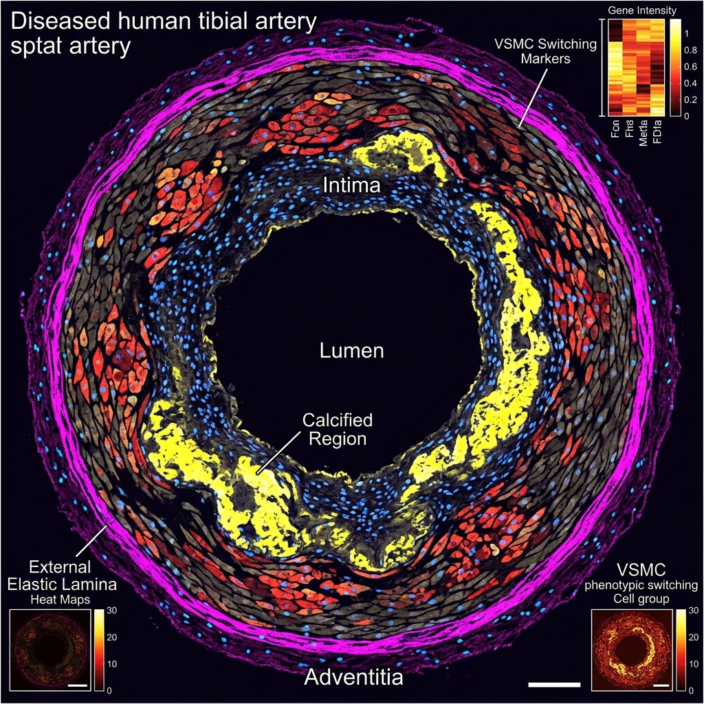
Xenium spatial transcriptomics of tibial artery
Xenium Spatial Omics

Bilayered Medial Architecture

Using the 10x Genomics Xenium platform to analyze human tibial vascular specimens, revealing a bilayered medial architecture with distinct inner and outer layers expressing unique gene signatures relevant to PAD.

Arterial calcification and VSMC phenotypic switching
Translational

Arterial Calcification in PAD

Studying the molecular processes driving arterial calcification in below-knee vascular segments, identifying structural changes that impair blood flow and threaten limb salvage in PAD patients.


Lab News & Announcements

Recent grants, awards, clinical milestones, and research highlights from the Vascular Media Lab.

Loading News & Updates...


Meet the Team

A multidisciplinary group of scientists passionate about understanding vascular biology at the molecular level.

Loading Team Profiles...

Past team members will appear here.


Publications

Our peer-reviewed contributions to understanding vascular biology and spatial transcriptomics.

Submitted Manuscript

Cellular-Resolution Spatial Transcriptomics Reveals Laminar VSMC Phenotypic Remodeling and a Hypoxic Medial Core in Human Thoracic Aortic Dissection

Holzem KM, et al.

Submitted (2026)

2025

Xenium Spatial Transcriptomic Analysis Reveals Bilayered Medial Architecture in Human Tibial Arteries

Holzem KM, et al.

Arteriosclerosis, Thrombosis, and Vascular Biology (AHA Scientific Sessions Abstract)

View Abstract

Understanding Aortic Dissection

This educational resource outlines critical safety symptoms, anatomical definitions, and diagnostic options for patients and families.

ðŸšĻ Call 911 Immediately

Sudden, severe chest, back, or abdominal pain — often described as ripping, tearing, or stabbing — is a sign of acute aortic dissection. This is an immediate medical emergency. Do not drive yourself; call 911 immediately.

What is a Type A Dissection?

An intimal flap tear that starts in the ascending aorta, allowing high-pressure blood flow to rip apart and split the middle (medial) tissue layers of the vessel wall. This requires emergency cardiac surgery.

Connective Tissue Risks

Certain genetic traits and connective tissue conditions like Marfan, Loeys-Dietz, and vascular Ehlers-Danlos syndromes increase susceptibility to arterial media tears and aneurysms.

Clinical Consultations

For surgical planning, medical details, and diagnostic tracking, view official medical program information at the WashU Medicine Aortic Center.


Contact Us

Interested in our research or potential collaboration? We'd love to hear from you. Reach out and we'll get back to you as soon as possible.

📍

Location

Washington University in St. Louis
St. Louis, MO 63130

✉ïļ
🏛ïļ

Department

Department of Surgery
Division of Vascular Surgery